14 Abdomen CT
Reference:
- CT Abdomen & Pelvis (YouTube) [1]
14.1 Steps
General axial scan
Free intraperitoneal gas or fluid ?
Gas: (use wide window) see whether gas in bowel ?
Fluid: see free fluid in dependent areas (hepatorenal, perihepatic, pelvis, etc.)
(Scroll down): Assess Organs
- Liver / GB: focal lesion, duct dilatation
- Pancreas: duct dilatation
- Spleen
- Adrenals
- Kidneys: hydronephrosis, follow ureter down to..
- Bladder:
(Scroll up): Glutter
- Lt paracolic glutter: greater omentum
- Rt paracolic glutter: morrison pouch, subphrenic space
Bowel
- Esophagus → Stomach
- Small bowel: (duodenum 1st - 4th) left → right “sweep”
- Colon:
- anus → rectum → sigmoid
- desc.colon → splenic.flex → TV → hep.flex → asc.colon
- cecum → terminal illeum
- Appendix
14.2 Esophagus
14.3 Bowel
- Diameter: small bowel <
3 cm, large bowel <6 cm, cecum <9 cm
Warning
Characteristic is more important than size, e.g., proximal distend, distal collapse, with transition zone suggests gut obstruction.
14.4 Biliary
- CBD: diameter <
6 mm(if age > 60 yr, can dilate 1 mm / yrs)
14.5 Pancreas
- Pancreatic duct diameter <
3 mm
14.6 Spleen
- Max diameter (axial or coronal) <
12 cm
14.7 Adrenal gland
Thickness <
1 cmConcave margins (look for convexity, e.g., nodules)
14.8 Renal
14.8.1 Renal protocol CT
Corticomedullary phase: mostly cortex enhanced
Nephrographic phase: mostly kidney parenchyma enhanced
Delayed phase: see collecting systems
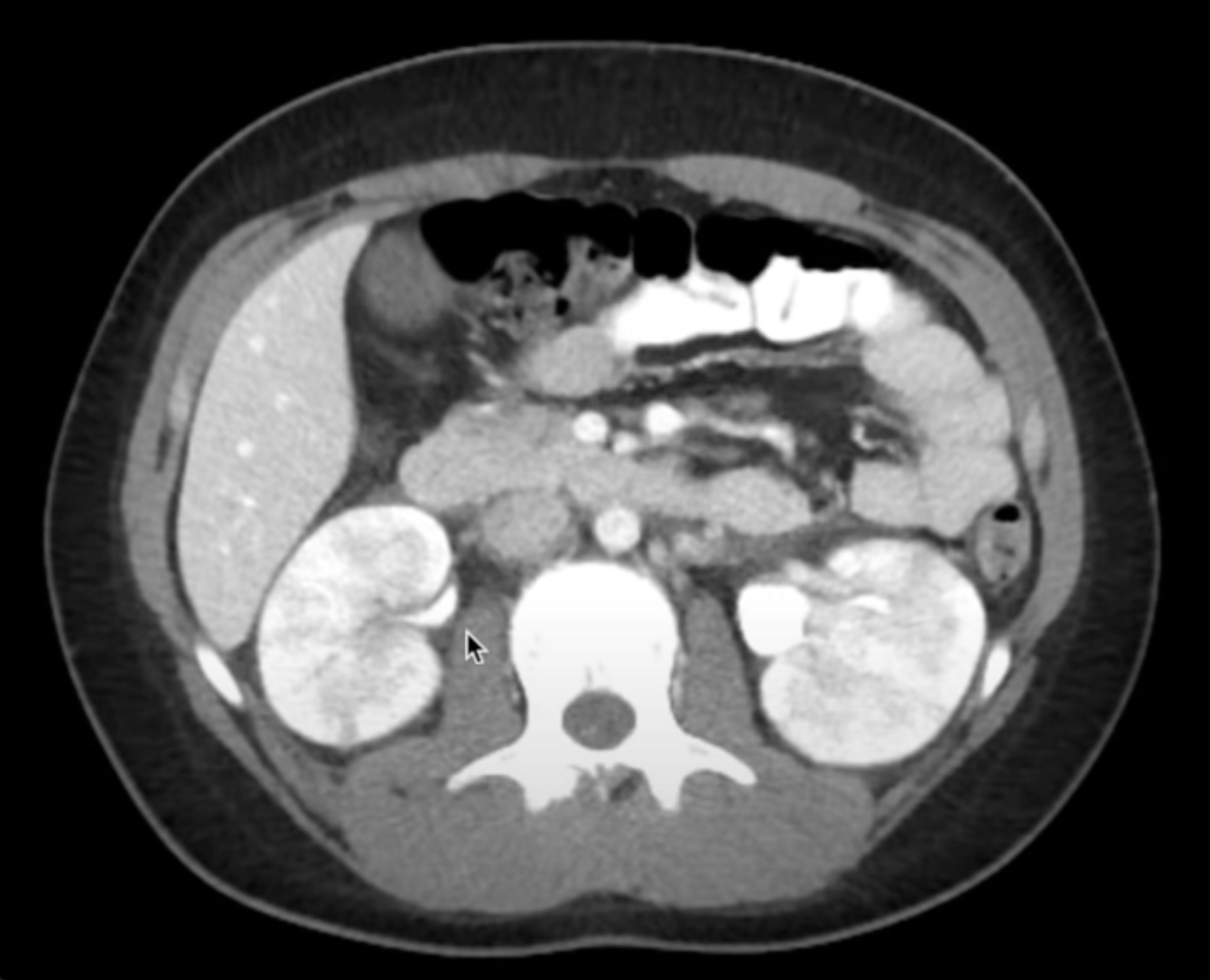
14.8.2 Pyelonephritis
See Striated nephrogram Figure 14.2
14.8.3 Hydroureter
- Ureter diameter >
3 mm(ref)
14.9 Lymphnodes (Abdomen)
14.9.1 Normal LN
Diameter: approx. < 1 cm in short axis, oval-shaped
14.9.2 Abnormal LN
- Shape: Rounded
- Heterogeneous / Cystic change

